|
ICP is determined by the volume
of the various intracranial components. Normal ICP
is approximately 10 mm Hg. Because the cranium has a
fixed volume, an increased volume of any
intracranial component must be compensated for by a
decrease in the volume of another component.
Otherwise, the pressure in the cranium will
increase.
There are three major intracranial components.
Brain tissue represents 80% to 85% of the
intracranial volume and is composed of a cellular
component that includes the neurons and glia, and an
extracellular component consisting of the
interstitial fluid.
The CSF volume accounts for 7% to 10% of the
intracranial volume.
The cerebral blood volume accounts for 5% to 8% of
the intracranial volume and includes the blood in
the vascular space.
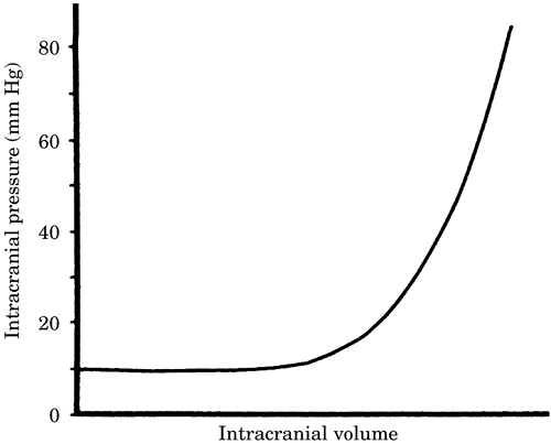
There is an elastance or compliance to the
components in the cranium such that a small increase
in the volume of one component does not cause an
increase in pressure. Once this compliance is
exhausted, small increases in volume lead to large
increases in ICP (Figure 1).
Increases in cranial volume can be caused by:
Increases in CSF volume due to blockage of the
circulation or absorption of CSF.
Increased cerebral blood volume due to
vasodilatation (intravascular) or hematoma
(extravascular).
Increased brain tissue volume due to a tumor or
edema.
Brain edema: Brain edema is classified as cytotoxic
or vasogenic and can increase ICP.
Cytotoxic edema is due to swelling of the neuronal
and/or glial cellular component and is frequently
the result of cerebral ischemia or trauma.
Vasogenic edema is caused by a breakdown of the BBB.
The resultant extra-vascularization of protein
increases interstitial water due to an increase in
osmotic equivalents in the extravascular space.

Figure 1. Idealized Intracranial pressure-volume
curve.
ICP pathophysiology. An increase in ICP can have
severe pathophysiologic consequences.
CPP can be calculated by subtracting ICP from mean
arterial pressure (MAP) (i.e., CPP = MAP - ICP).
As ICP increases, CPP decreases. This leads to
cerebral ischemia. Normally cerebral ischemia leads
to the Cushing reflex, which increases MAP. However,
this can compensate only up to a certain point
beyond which CPP will fall further, leading to
severe ischemia, coma, and death if ICP is
uncontrolled.
Increased ICP can also cause herniation of the
brain, which can lead to rapid neurologic
deterioration and death.
|